Why Fiber Optics?
Fiber optics use total internal reflection to guide photons through ultra‑pure glass cores, delivering light and data into places electrical conductors simply cannot. Because light is immune to electromagnetic interference and glass is non‑conductive, fiber tools are safe beside MRI magnets, electrosurgical units, and radiofrequency ablation probes.
Top Benefits in Medicine
Benefit | Impact in the OR/Clinic |
|---|---|
High Bandwidth | HD video and multispectral imaging on a single strand |
Immunity to EMI | Zero signal loss near strong magnetic fields |
Miniaturization | Catheters ≤ 1 mm OD for pediatric and neuro procedures |
Biocompatibility | Inert silica glass meets USP Class VI |
Remote Energy Delivery | Accurate laser therapy deep inside the body |
Clinical Application Deep‑Dive
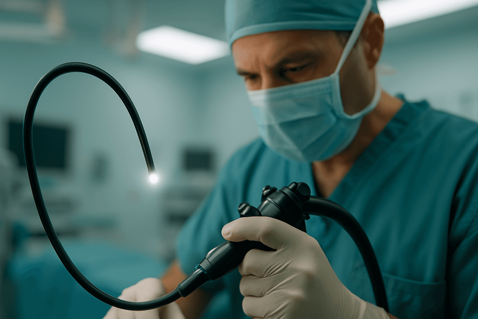
Endoscopy & Arthroscopy
White‑light illumination + fiber image bundles give surgeons live HD visuals through keyhole incisions.
New chip‑on‑tip scopes combine single‑mode illumination fibers with CMOS sensors for 4K imagery down bronchial branches and GI tracts.
Laser Surgery
Multimode fibers route kilowatt‑class IR lasers for tumor ablation, lithotripsy, and photocoagulation.
Polymer‑clad silica withstands > 200 °C at the distal tip, ensuring thermal stability.
Optical Coherence Tomography (OCT)
Uses interferometry in single‑mode fiber to create 5 µm axial‑resolution cross‑sections of retina, coronary plaques, and skin layers.
Swept‑source lasers at 1,300 nm penetrate deeper with less scattering.
Catheter Guidance & IV Placement
Side‑firing fibers project structured light onto vessel walls; CMOS capture returns real‑time depth maps.
First‑stick success often exceeds 95 %, slashing complications and supply costs.
In‑Vivo Fiber Sensors
Fiber Bragg gratings (FBGs) embedded along catheter shafts monitor temperature, pressure, and strain.
Safe inside MRI due to non‑metallic construction.
Engineering & Design Factors
Parameter | Typical Spec | Why It Matters |
Core Diameter | 5 – 9 µm (SM), 50 µm (MM) | Controls mode structure and power delivery |
Numerical Aperture | 0.12 – 0.22 | Higher NA collects more light but increases dispersion |
Bend Radius | ≤ 5 mm with < 0.5 dB loss | Critical for routing through tight anatomical curves |
Coating & Buffer | Polyimide, Parylene | Survives autoclave and ETO sterilization |
Connectors | Luer‑lock, FC/PC, Keyed ST | Maintain sterility and repeatable alignment |
Mini‑Tip: FSI’s draw‑tower lets us tweak dopants for low‑OH attenuation at UV wavelengths (270 – 400 nm) critical for fluorescence imaging.
Sterilization & Regulatory Pathways
Steam Autoclave (134 °C, 2 bar) – Most durable glass and polyimide coats survive 1,000+ cycles.
Plasma Sterilization (H₂O₂) – Ideal for heat‑sensitive polymer endcaps.
Ethylene Oxide (ETO) – Deep penetration for long lumens, though requires out‑gassing time.
Reg Note: Fiber devices touching sterile tissue are typically Class II (US FDA) and Class IIa/IIb (EU MDR). FSI supplies full material traceability, biocomp test data, and validated cleaning/sterilization protocols to streamline 510(k) or CE submissions.
Case Studies
A. Pediatric Cardiology – Real‑Time OCT Catheter
A Midwest children’s hospital integrated a 0.9 mm OCT catheter with FSI’s bend‑insensitive single‑mode fiber. Surgeons visualized congenital heart defects in situ, reducing cardiac bypass times by 18 %.
B. Oncology – Multispectral Endoscope
A UK cancer center deployed a dual‑fiber system: one fiber delivered UV excitation while a return fiber captured fluorescence. Detection of small bowel tumors improved by 22 % in early trials.
Emerging Research
Photonic Neural Probes: 10,000‑core fibers stimulate and read neuronal circuits with millisecond precision.
Power‑over‑Fiber Implants: Optical power + data eliminate batteries in pacemakers and drug pumps.
AI‑Enhanced Endoscopy: On‑probe GPUs analyze images in real time, flagging polyps under 5 mm.
FSI Advantage
Custom Glass Recipes via in‑house draw‑tower (low‑OH, radiation hard, specialty dopants).
Micro‑Assembly: Terminations down to 400 µm ID ferrules.
Regulatory Support: DHF documentation, risk analysis, and biocompatibility data packages.
**Need a prototype or a 10,000‑unit production run? **Talk with FSI’s medical engineering team →